
 Back
BackX-Ray Imaging
X-ray imaging is a medical imaging technique that uses X-rays to view the internal structure of the body
03/2026
BackX-ray imaging is a medical imaging technique that uses X-rays to view the internal structure of the body
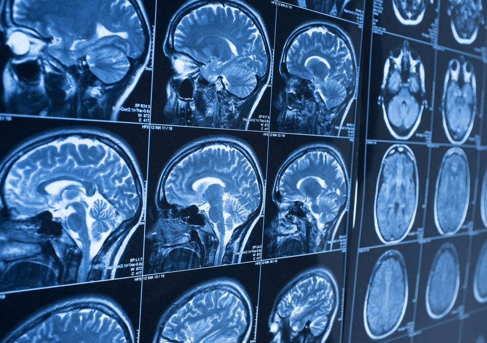
X-ray imaging is a medical imaging technique that uses ionizing radiation to view the inside of the body. It is one of the most common and widely used diagnostic tools in medicine. X-rays can help detect fractures, tumors, and other abnormalities. The process involves exposing the body to a small dose of ionizing radiation, which passes through the body and is absorbed by different tissues unequally. This creates a contrast that can be captured on film or digitally. But how does it work?
The standard device for generating X-rays is the vacuum tube. The physical process involves three main steps:
Higher kV increases electron speed and X-ray energy (penetration power).
Higher mA increases the number of electrons, producing more X-ray photons.
While the previous interactive model explains the fundamental physics, a real modern X-ray tube is slightly more complex. The most significant difference is the rotating anode.
Remember that 99% of the kinetic energy from the electrons is converted into heat. If the electron beam continuously hit the exact same spot on a stationary target, the tungsten would quickly melt.
To solve this, the anode is designed as a large beveled disk attached to a rotor. As the disk spins at high speeds (often 3,000 to 10,000 RPM), the electron beam strikes a constantly moving circular path called the focal track. This spreads the huge amount of heat over a much larger surface area, allowing for higher energy exposures without destroying the tube.
Beyond the X-ray tube itself, the complete instrumentation includes essential components placed immediately after the tube window to condition the X-ray beam before it reaches the patient.
Once emitted, X-rays pass through the patient. The image is formed due to beam attenuation according to the Beer-Lambert law:
Where is the final intensity, the initial intensity, the attenuation coefficient (which depends on tissue density), and the thickness traversed.
Modern imaging has evolved using now digital sensors. There are 2 main ways we capture X-rays today:
The newest revolution in CT Scanning. These detectors count every single photon instead of measuring an average "cloud" of energy. This allows for precised detail at a much lower dose.
X-rays hit the Scintillator, creating a burst of visible light (scattering). This light is then converted into electrical charge by the Photodiode, which is read by the TFT array. The light scattering slightly reduces image sharpness.
Now that we have a basic understanding of X-ray production interaction and detection, let's explore how these rays interact with different tissues in the body.
Discovered by Wilhelm Röntgen in 1895 (who took the first radiograph of his wife's hand), X-rays pass through the body and are absorbed differently depending on tissue density.
Dense bones appear white because they absorb most of the rays, preventing them from reaching the detector. Conversely, soft tissues and air allow rays to pass through, appearing in shades of gray or black.
Beyond standard static radiography, there are other types of specialized imaging systems. One of them is the C-Arm, a mobile device named for its "C"-shaped arm. This design is on purpose, because it allows the device to be easily moved around the operating table, providing imaging from almost any angle without the need to move the patient during surgery.
Commonly used in orthopedic, vascular, and digestive surgeries, the C-Arm transforms static X-rays into Fluoroscopy, a continuous "live video" feed at up to 30 frames per second. This provides surgeons with a real-time view inside the body, acting as a guide during complex procedures.
Additionally, by using contrast agents to "light up" the vascular system, doctors can perform minimally invasive surgeries through a tiny incision rather than opening the chest. These agents are highly visible because they contain elements with a high atomic number (like Iodine) that have denser electron shells and a stronger nuclear pull. This creates a much larger target for X-ray photons to hit, exponentially increasing the probability of absorption (Photoelectric Effect) compared to surrounding soft tissues.
In live fluoroscopy, we switch to Negative Mode, where dense objects appear black, because it is significantly easier for the human eye to track thin surgical tools like wires and stents against a lighter background.
Martin Berger, Qiao Yang, and Andreas Maier. (2018)
Medical Imaging Systems: An Introductory Guide [Internet]. Cham (CH): Springer; 2018. Chapter 7.Samuel J. Ling, Jeff Sanny, William Moebs (2016)
OpenStax - University Physics Volume 3 : 8.5 Atomic Spectra and X-rays